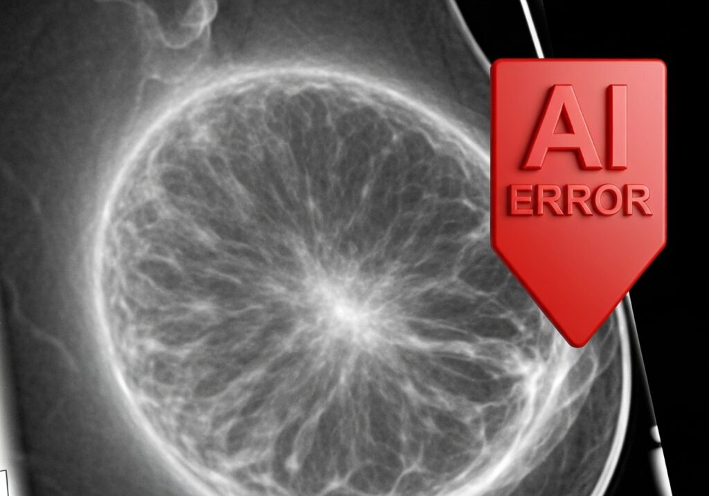
NAVIGATING THE NUANCES OF ARTIFICIAL INTELLIGENCE IN MAMMOGRAPHY: UNDERSTANDING FALSE NEGATIVES
The integration of artificial intelligence (AI) into diagnostic imaging, particularly mammography, has ushered in a new era of breast cancer screening with immense promise for enhanced accuracy and efficiency. AI algorithms are designed to assist radiologists in detecting subtle abnormalities, reducing workload, and potentially improving patient outcomes. However, like any sophisticated technology, AI is not infallible. A recent study published in Radiology sheds critical light on the factors contributing to false negatives in AI-powered mammography analysis, offering invaluable insights for both clinical practice and future AI development. This article delves into the findings of this pivotal research, exploring the key characteristics that lead AI software to miss invasive breast cancers and discussing the broader implications for the evolving landscape of breast imaging.
THE PROMISE AND PITFALLS OF AI IN BREAST IMAGING
Artificial intelligence, particularly in the form of deep learning, has rapidly advanced in its ability to analyze complex medical images. In mammography, AI systems are trained on vast datasets of breast images to identify patterns indicative of malignancy. The primary goals are to:
- Enhance Detection Rates: By identifying subtle cancers that might be overlooked by the human eye, especially in challenging cases.
- Reduce Radiologist Workload: By prioritizing studies or flagging suspicious areas, allowing radiologists to focus on complex cases.
- Improve Efficiency: Speeding up the interpretation process.
- Standardize Interpretation: Reducing inter-reader variability.
Despite these compelling advantages, the critical question remains: where does AI fall short? Understanding its limitations is as important as recognizing its strengths, especially when patient lives are at stake. False negatives, where cancer is present but not detected, represent a significant concern in screening programs, as they can delay diagnosis and treatment.
UNVEILING THE STUDY’S FINDINGS: A CLOSER LOOK AT AI’S PERFORMANCE
The retrospective study, conducted by Ok Hee Woo, M.D., and her team at Korea University Guro Hospital in Seoul, analyzed the performance of a specific AI software (Lunit Insight MMG, version 1.1.7.3, Lunit) on mammograms from 1,082 women diagnosed with 1,097 invasive breast cancers. The findings were stark and highly informative:
The AI software demonstrated a 14 percent false-negative rate, missing 154 out of 1,097 invasive breast cancers. Crucially, 61.7 percent of these missed cases were deemed “actionable,” meaning they represented clinically significant cancers that should have been detected. This highlights that these were not merely borderline cases but instances where the AI failed to identify actual malignancies.
The researchers then meticulously investigated the characteristics of these AI-missed cancers, revealing several key factors that contribute to these detection failures.
KEY FACTORS CONTRIBUTING TO AI FALSE NEGATIVES
The study identified three primary factors that significantly correlated with AI-missed breast cancers, alongside other contributing patient and tumor characteristics.
1. BREAST DENSITY: A PERSISTENT CHALLENGE
One of the most prominent findings was the strong association between dense breast tissue and AI false negatives.
- Prevalence: 59 percent of actionable AI-missed breast cancers occurred in women with dense breasts.
- Why it’s challenging: Dense breast tissue, which consists of more glandular and fibrous tissue than fatty tissue, appears white on mammograms. Cancerous lesions also appear white, making it incredibly difficult for both human radiologists and AI algorithms to distinguish between normal dense tissue and a cancerous mass. This “masking effect” is a well-known hurdle in mammography. For AI, the increased signal complexity and reduced contrast between normal and abnormal tissue in dense breasts can hinder its ability to identify subtle cancerous patterns.
2. NON-MAMMARY ZONE LOCATIONS: BEYOND THE USUAL SUSPECTS
Another significant contributor to AI misses was the location of the tumor.
- Prevalence: 23 percent of AI-missed cases involved cancers located in “non-mammary zones.”
- What are non-mammary zones? These typically refer to areas outside the main glandular breast tissue, such as the far axilla (armpit region), pectoral muscle region, or very peripheral areas of the breast. These locations can be challenging for AI due to:
- Variability in Imaging: These areas might not always be perfectly captured or optimized in standard mammographic views.
- Limited Training Data: AI models are often trained predominantly on cancers within the central breast parenchyma, potentially leading to less robust performance in less common anatomical locations.
- Anatomical Overlap: Structures like the pectoral muscle or ribs can obscure lesions in these peripheral areas.
3. TUMOR CHARACTERISTICS: SIZE AND BIOLOGICAL BEHAVIOR
The study also revealed crucial differences in the characteristics of AI-missed cancers compared to AI-detected ones.
- Tumor Size: Counterintuitively, AI-missed cancers had a higher incidence of tumors ≤ 2 cm (81.8 percent vs. 61 percent for AI-detected cancers). While one might expect AI to miss smaller, more subtle lesions, the data suggests that for a significant proportion of AI-missed cases, the tumors were not necessarily microscopic but fell within a size range that AI struggled with for other reasons. This could be due to their morphology, location, or surrounding tissue.
- Histologic Grade and Lymph Node Involvement: AI-missed cancers were associated with a lower histologic grade and fewer lymph node metastases (18.2 percent vs. 31.1 percent for AI-detected). This suggests that AI might be less adept at identifying less aggressive or earlier-stage cancers, which often present with more subtle imaging features.
- Molecular Subtypes: The AI system showed varying false-negative rates across different molecular subtypes of breast cancer:
- Luminal: Highest FNR at 17.2 percent. Luminal cancers are the most common type and often present as less aggressive masses.
- Triple-Negative: FNR of 14.5 percent. Triple-negative cancers are often aggressive but can be difficult to detect if they don’t form a distinct mass or are obscured.
- HER2-Enriched: Lowest FNR at 9 percent. This aligns with prior research suggesting AI’s proficiency in detecting HER2-positive tumors, often due to their association with microcalcifications, which AI algorithms are generally very good at identifying.
- BI-RADS Category: AI-missed cancers frequently received BI-RADS category 4 interpretations, indicating suspicious but not definitively malignant findings. This suggests that even when a human reader identified a suspicious area, the AI’s abnormality score for that lesion remained low.
4. PATIENT AGE: A DEMOGRAPHIC FACTOR
The study found that women with AI-missed invasive breast cancers were, on average, over five years younger (mean age of 49.7 years) compared to those with AI-detected cancers (mean age of 55.1 years). Younger women often have denser breast tissue, which could be a contributing factor to the AI’s reduced sensitivity in this demographic.
IMPLICATIONS FOR CLINICAL PRACTICE AND THE ROLE OF HUMAN OVERSIGHT
These findings have profound implications for how AI is integrated into clinical mammography workflows:
- AI as an Aid, Not a Replacement: The study strongly reinforces the concept that AI should serve as an assistive tool, augmenting human interpretation rather than replacing it. Radiologists remain indispensable for reviewing cases flagged by AI and, crucially, for scrutinizing cases where AI might underperform.
- Awareness of AI’s Blind Spots: Radiologists need to be keenly aware of the specific scenarios where current AI algorithms are more prone to false negatives. This includes patients with dense breasts, lesions in non-mammary zones, and certain tumor characteristics.
- Emphasis on Multimodality Imaging: For high-risk patients or those with dense breasts, supplemental imaging modalities like ultrasound or MRI may be even more critical when AI mammography is employed. These modalities can often overcome the masking effects of dense tissue.
- Continued Training and Validation: AI algorithms must continue to be trained on diverse datasets that include challenging cases, particularly those involving dense breasts and peripheral lesions. Future generations of AI should aim to specifically address these identified limitations.
ADDRESSING LIMITATIONS AND CHARTING THE PATH FORWARD
While invaluable, the study acknowledged several limitations that warrant consideration:
- Retrospective Design: This type of study looks back at existing data, which can introduce biases. Prospective studies, designed specifically to evaluate AI performance in real-time screening settings, would provide stronger evidence.
- Single AI Software: The study used only one specific AI software (Lunit Insight MMG). Performance can vary significantly between different AI models and vendors. Generalizing these findings to all AI mammography systems should be done with caution.
- Cohort Composition: The study cohort consisted only of women with confirmed invasive breast cancer. This means the false-positive rate of the AI (identifying something as cancer when it’s not) was not the focus, and the population had a higher prevalence of dense breasts and BI-RADS category 5 lesions than a typical screening population. Future research should evaluate AI in broader, more representative screening cohorts.
Despite these limitations, the study provides a critical framework for understanding and improving AI’s role in breast imaging. Future research should focus on:
- Developing AI algorithms specifically optimized for dense breasts and atypical tumor locations.
- Integrating multi-modal imaging data (mammography, ultrasound, MRI) into AI analysis for a more comprehensive assessment.
- Conducting large-scale, prospective studies with diverse patient populations to validate AI performance in real-world screening scenarios.
- Exploring how AI can better interpret subtle features that characterize less aggressive or earlier-stage cancers, which were often missed in this study.
CONCLUSION: EVOLVING WITH INTELLIGENCE AND INSIGHT
The new study on false negatives in AI mammography analysis serves as an essential reminder of both the immense potential and the inherent complexities of artificial intelligence in healthcare. While AI offers unparalleled tools to enhance breast cancer detection, its limitations, particularly concerning dense breasts, non-mammary zone lesions, and certain tumor characteristics, must be thoroughly understood and addressed. This research underscores the indispensable role of the human radiologist in the diagnostic process—not merely as a supervisor, but as a critical interpreter who can leverage AI’s strengths while compensating for its weaknesses. As AI technology continues to evolve, ongoing research and a collaborative approach between developers, clinicians, and patients will be vital to ensure that these powerful tools truly optimize breast cancer screening and improve outcomes for all.

